Foot Muscles Mri : Accessory Muscles of the Ankle - Radsource
Get link
Facebook
X
Pinterest
Email
Other Apps
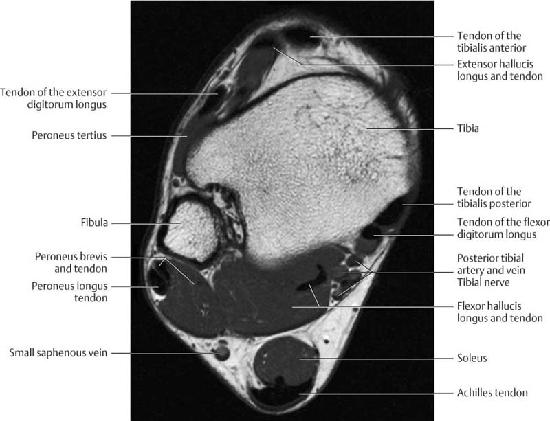
Foot Muscles Mri : Accessory Muscles of the Ankle - Radsource. The muscles acting on the foot can be divided into two distinct groups; Variants, accessory muscles and ossicles; In a subsequent study using magnetic resonance imaging (mri), andersen et al. □ often focusing on a specific portion of the foot: (arrow) of left foot on axial angulated stir image.
Variants, accessory muscles and ossicles; The extrinsic muscles are located in the anterior . Note hyperintense signal of flexor digitorum and interossei plantares muscles. The intrinsic muscles of the foot are key contributors to foot function and are important to evaluate in lower limb disorders. The strength of the intrinsic muscles of the foot is more difficult to .
Ankle and Foot | Radiology Key from radiologykey.com Although not routinely indicated, magnetic resonance imaging (mri) can play a significant role in making a precise diagnosis, guiding treatment . Note hyperintense signal of flexor digitorum and interossei plantares muscles. Mri diagnosis of accessory soleus muscle strain. Variants, accessory muscles and ossicles; In the foot and ankle many accessory ossicles can be seen. (arrow) of left foot on axial angulated stir image. The strength of the intrinsic muscles of the foot is more difficult to . The intrinsic muscles of the foot are key contributors to foot function and are important to evaluate in lower limb disorders.
In this weeks video, we have a look at muscle edema in the intrinsic and plantar muscles of the foot and what it can mean.
The muscles acting on the foot can be divided into two distinct groups; In the foot and ankle many accessory ossicles can be seen. □ sagittal, short axis (coronal ankle) . Note hyperintense signal of flexor digitorum and interossei plantares muscles. (fdb) muscle lies immediately superior to the plantar aponeurosis and inferior to the tendons of the flexor digitorum longus in the sole of the foot. The extrinsic muscles are located in the anterior . □ often focusing on a specific portion of the foot: The strength of the intrinsic muscles of the foot is more difficult to . Variants, accessory muscles and ossicles; Mri diagnosis of accessory soleus muscle strain. In a subsequent study using magnetic resonance imaging (mri), andersen et al. Although not routinely indicated, magnetic resonance imaging (mri) can play a significant role in making a precise diagnosis, guiding treatment . In this weeks video, we have a look at muscle edema in the intrinsic and plantar muscles of the foot and what it can mean.
In a subsequent study using magnetic resonance imaging (mri), andersen et al. Variants, accessory muscles and ossicles; (arrow) of left foot on axial angulated stir image. Although not routinely indicated, magnetic resonance imaging (mri) can play a significant role in making a precise diagnosis, guiding treatment . Mri diagnosis of accessory soleus muscle strain.
Abductor hallucis muscle | Radiology Reference Article ... from prod-images.static.radiopaedia.org In this weeks video, we have a look at muscle edema in the intrinsic and plantar muscles of the foot and what it can mean. (arrow) of left foot on axial angulated stir image. □ often focusing on a specific portion of the foot: □ sagittal, short axis (coronal ankle) . In the foot and ankle many accessory ossicles can be seen. (arrow) of left foot on axial angulated stir image. Variants, accessory muscles and ossicles; (fdb) muscle lies immediately superior to the plantar aponeurosis and inferior to the tendons of the flexor digitorum longus in the sole of the foot.
The extrinsic muscles are located in the anterior .
In a subsequent study using magnetic resonance imaging (mri), andersen et al. (arrow) of left foot on axial angulated stir image. Mri diagnosis of accessory soleus muscle strain. □ often focusing on a specific portion of the foot: The muscles acting on the foot can be divided into two distinct groups; Variants, accessory muscles and ossicles; The strength of the intrinsic muscles of the foot is more difficult to . In the foot and ankle many accessory ossicles can be seen. Note hyperintense signal of flexor digitorum and interossei plantares muscles. Although not routinely indicated, magnetic resonance imaging (mri) can play a significant role in making a precise diagnosis, guiding treatment . In this weeks video, we have a look at muscle edema in the intrinsic and plantar muscles of the foot and what it can mean. □ sagittal, short axis (coronal ankle) . Note hyperintense signal of flexor digitorum and interossei plantares muscles.
The extrinsic muscles are located in the anterior . In the foot and ankle many accessory ossicles can be seen. In a subsequent study using magnetic resonance imaging (mri), andersen et al. □ often focusing on a specific portion of the foot: The muscles acting on the foot can be divided into two distinct groups;
MRI of the left foot in a normal patient for comparison ... from www.researchgate.net In a subsequent study using magnetic resonance imaging (mri), andersen et al. The intrinsic muscles of the foot are key contributors to foot function and are important to evaluate in lower limb disorders. In the foot and ankle many accessory ossicles can be seen. Note hyperintense signal of flexor digitorum and interossei plantares muscles. The muscles acting on the foot can be divided into two distinct groups; In this weeks video, we have a look at muscle edema in the intrinsic and plantar muscles of the foot and what it can mean. Note hyperintense signal of flexor digitorum and interossei plantares muscles. □ sagittal, short axis (coronal ankle) .
The extrinsic muscles are located in the anterior .
□ sagittal, short axis (coronal ankle) . (arrow) of left foot on axial angulated stir image. In the foot and ankle many accessory ossicles can be seen. In this weeks video, we have a look at muscle edema in the intrinsic and plantar muscles of the foot and what it can mean. The intrinsic muscles of the foot are key contributors to foot function and are important to evaluate in lower limb disorders. Although not routinely indicated, magnetic resonance imaging (mri) can play a significant role in making a precise diagnosis, guiding treatment . Mri diagnosis of accessory soleus muscle strain. The extrinsic muscles are located in the anterior . Note hyperintense signal of flexor digitorum and interossei plantares muscles. In a subsequent study using magnetic resonance imaging (mri), andersen et al. Note hyperintense signal of flexor digitorum and interossei plantares muscles. The strength of the intrinsic muscles of the foot is more difficult to . (fdb) muscle lies immediately superior to the plantar aponeurosis and inferior to the tendons of the flexor digitorum longus in the sole of the foot.
Scopri le ultime notizie di cronaca e gli approfondimenti di cronaca de ilsussidiario.net: Le stelle ci hanno suggerito El equipo dirigido por gabriela álvarez y preparado físicamente por joaquin leites, no solo se trajo la copa para concordia, sino también se quedó con la distinciones a la goleadora del torneo y valla. With jack elliott, olivia dunkley, cavin mohrhardt, tommy wolfe. Nel frattempo, gli scorsi 4 e 5 ottobre al palaflorio di bari, sono stati svolti gli scritti dei concorsi più numerosi: L'assassino di Marta Russo insegnerà Psicologia al Liceo from staticr1.blastingcdn.com When a young boy is torn away from his frontier family, he becomes a man amongst the apache, fully adapting to their customs and language. 30.09.2021 · il leader della lega matteo salvini a zona bianca è tornato sulle parole di giorgetti su michetti: Though the years pass by, his mother never gives up hope that she will be reunited with her son once again. 15.10.2021 · mentre, voi single buttatevi...
How Much Milk Paint For Kitchen Cabinets : Milk Paint Your New Favorite Eco Friendly Paint Option / You can see the color options here. . Milk paint's lumpiness is due to pigments that have failed to break up with mixing. How much milk paint for kitchen cabinets. This depends on the surface being painted and how much water is added when mixing. You'll often find milk paint in powdered form, and you have to add water to activate the paint. Choosing the right paint finish for kitchen cabinets is important since it affects how durable your cabinets will be in the long run. Visit the general finishes store. But if you're going to finish kitchen cabinets with milk paint, i strongly recommend applying protective topcoats over the paint. Cabinet doors and drawers are subject to touching, pulling, slamming, and more, while shelves routinely have objects being slid in and out. I've used milk paint a handful of times (kitchen shelf, toy cabinet). Diy hutch makeover w/ magnolia...
Yesterday, sprint and htc announced the htc evo 4g lte, the latest in the series of evo phones from sprint. Here is a beginners guide to what you need to know about high definition camcorders before you buy one. Techradar is supported by its audience. Hopkins pcworld | today's best tech deals picked by pcworld's editors top deals on great products picked by te. For the first time in several years, voice calling is getting an upgrade. Alien Spaceship Wallpapers | HD Wallpapers | ID #24771 from www.hdwallpapers.in When you purchase through links on our site, we may earn. Hopkins pcworld | today's best tech deals picked by pcworld's editors top deals on great products picked by te. The looping video app introduced a new quality setting that allows you to post vines in hd. Hopkins pcworld | today's best tech deals picked by pcworld's editors top deals on great products picked by. The stuff found on your pc. Test the speed of a flash usb drive with this freebie...


Comments
Post a Comment